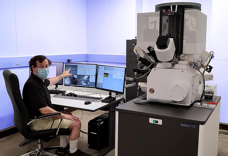
The Eyring Materials Center (EMC) proudly announces the addition of the Helios 5 UX dual beam, a Focused Ion Beam (FIB) Scanning Electron Microscope (SEM). This state-of-the-art instrument offers a significant improvement to our center’s electron microscopy capabilities. With its arrival, the Helios 5 UX provides the ability to produce exceptional sample lamella for transmission electron microscope (TEM) analysis. It can also generate sub-nanometer resolution images, previously not possible using any ASU SEM. The array of options available on the Helios5 makes it a powerful tool to address a wide variety of research challenges.
Electron microscopy has been part of ASU research since 1962, when the first transmission electron microscope (TEM) was installed. Today, the Eyring Materials Center is equipped with an array of TEMs used to study materials, including life science samples, at scales ranging from micro to atomic. Such studies require sample preparation, which can often be challenging especially in cases when a very specific location within the sample is to be studied.
To address such challenges, 16 years ago, the EMC installed ASU’s first dual beam FIB/SEM (the Nova200 Nanolab from FEI). The instrument is able to use gallium ions to cut through the material while imaging the sample with both the ions and electrons. With this tool researchers are able to not only cut through microscopic sample to observe the inside but also produce very thin – often less than 100nm lamella from the samples from a specific area, pluck them out using a micromanipulator and finally mount them on a TEM grid. With the advances in resolution and decrease in beam energy from standard TEM to the aberration correction TEM, the quality of the TEM samples from the Nova200 are often a limiting factor in the sample analysis. While still often used, the older technology of the Nova 200 often produces significant surface damage, requiring the sample to be thicker, which degrades the image quality and resolution and can also possibly affect the interpretation of the results.
In 2018, ASU’s Knowledge Enterprise awarded the EMC funding to purchase the new helios. The instrument, like many electron microscopes, has extremely stringent room requirements such as vibrational, electromagnetic field and temperature variation limitations. With Knowledge Enterprise’s outstanding support, the center retrofitted an existing TEM lab, in the basement of the Physical Science B-wing, into the perfect space for the new instrument. This summer, service engineers from Thermofisher Scientific completed the install of the new Helios 5 UX.
The advances in both hardware and software, now available with the Helios5, makes it uniquely valuable to a range of researchers. The new generation hardware provides excellent high-resolution SEM images even with challenging samples. The operator can use the ion beam to produce outstanding TEM samples with minimal damage thanks to the very low voltage capabilities. Using the Scanning TEM detector, the sample can then be imaged to ensure sample quality and provide preliminary data before moving he sample to one of our aberration-corrected TEMs. One can also slice the sample and image it after each pass in order to generate a 3D reconstruction of the sample. Beyond the imaging and cutting capabilities, the multiple gas injections systems (GIS) also allow the operator to deposit materials, such as carbon, tungsten or platinum to protect and attach samples, but also oxide insulating layers to do more advanced circuit modifications.
The upcoming addition of the cryogenic stage will further expand the scientific reach of the Helios. The near liquid nitrogen sample temperature will reduce beam-induced damage. The low temperature will also enable FIB milling of a vitrified frozen sample, to prepare it for TEM analyses such as cryotomography.
The Helios 5 UX, with its vast applications, is a remarkable addition to the center. The EMC team is eager to assist with the analytical needs of the research community using the new Helios5UX. You can contact them at emc@asu.edu. More information on the instrument can be found here (https://cores.research.asu.edu/node/402/)

