
Welcome to the ASU Core Facilities Newsletter. We are ready to support all your research goals. Please follow our LinkedIn page for additional resources and community information.
Highlight: Histology
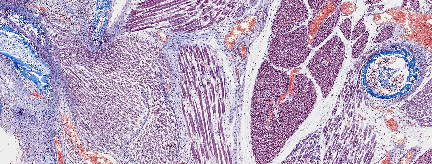
Histology is the study of the microanatomy of cells, tissues and organs as seen through a microscope. It examines the correlation between structure and function.
Histology examines tissue organization from cellular to organ levels. Removed tissue is processed to maintain cell structure, thinly sliced using a microtome or cryostat and placed on glass slides. Staining with dyes enhances contrast, allowing detailed study under an optical microscope.
Introducing our New Histology Core
The new Histology Core automates almost every process used in histology, from the initial tissue processing (including fixation, dehydration, clearing and paraffin infiltration) to paraffin embedding. The Histology Core also offers cassette and slide labeling and slide staining. Automation ensures that results will be reliable, consistent and reproducible.
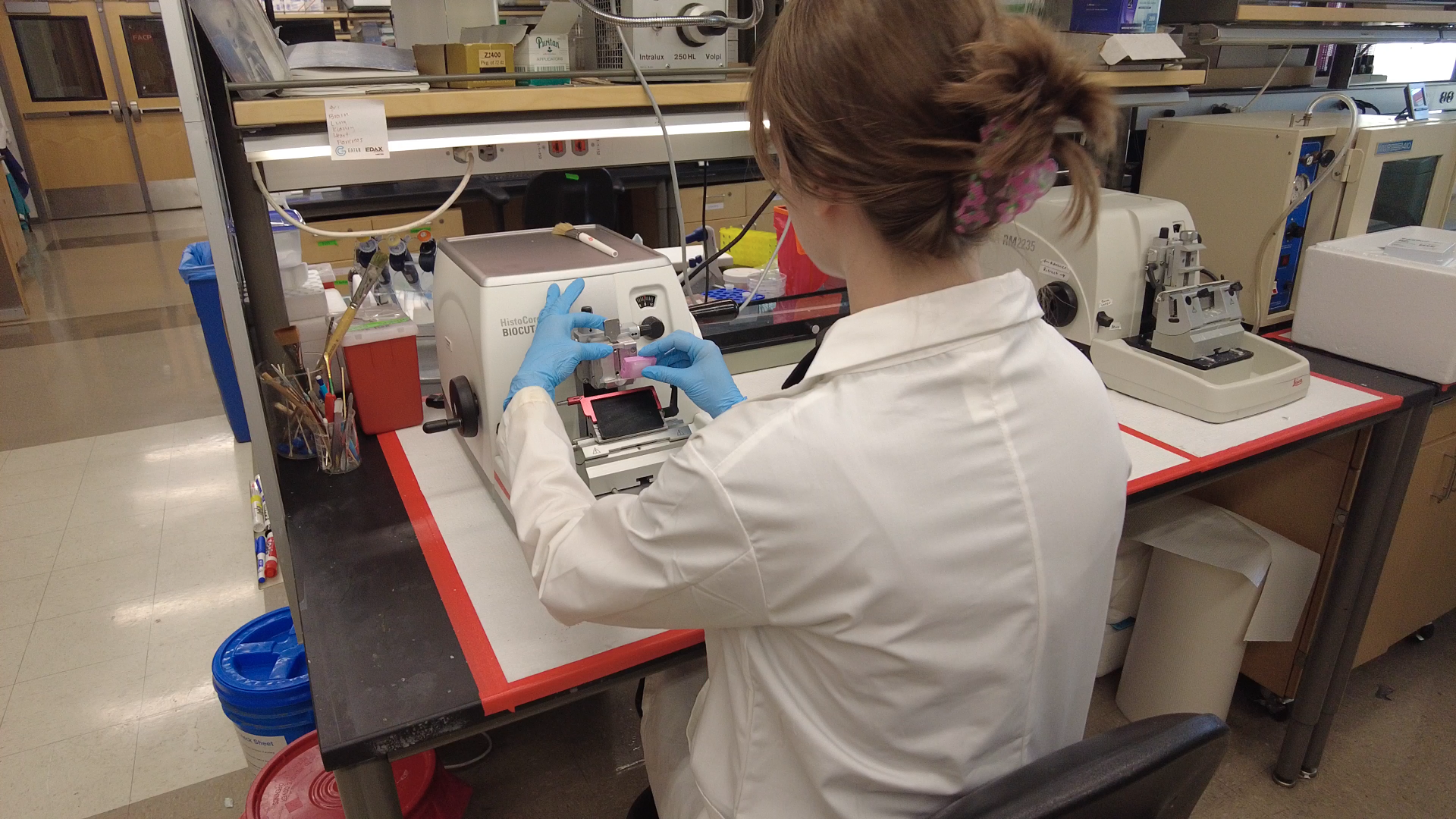
A new microtome and cryostat have been installed to ensure precise and consistent sections of tissue.
Histology Equipment
Leica HistoCore PEARL Automated Tissue Processor
The HistoCore PEARL automatically prepares tissue samples for laboratory testing by fixing, dehydrating, clearing and infiltrating them with paraffin. This fully enclosed tissue processor offers optimized fume protection for safe and reliable processing with up to 200 cassettes per run capacity. Pre-validated processing protocols can be adjusted to fit the unique needs of each individual tissue or specimen type.
This tissue processor is an asset to ASU research since it can replace and improve the results of manual processing due to the automated processors ability to provide consistent and reproducible results with a faster turnaround time.
Leica HistoCore Arcadia Embedding Center
Composed of the Arcadia H heated embedding workstation and the Arcadia C cold plate allows for simple operation and precise control; resulting in a smooth workflow, reliability and speed of your embedding work.
The HistoCore Arcadia C is a cold plate holding up to 65 cassettes on its large working surface. Cooling efficiency is important, so the cold plate was designed with an environment adaptive control module to make sure the operating temperature is stabilized at -6°C.
The HistoCore Arcadia Embedding Center is an asset to ASU research that increases the reliability and speed of the embedding process, resulting in consistent and reproducible results.
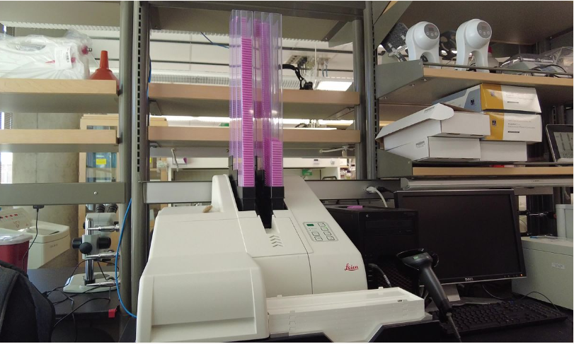
Leica ST5010 Autostainer XL
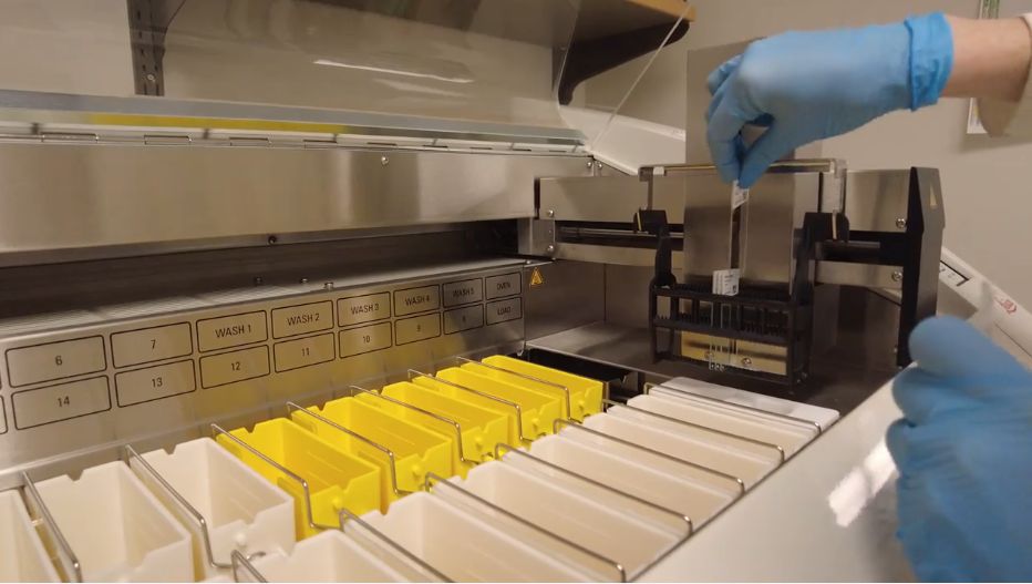
This automated slide stainer accommodates up to 11 racks of 30 slides each. The ability to store and execute 15 different user-defined staining protocols provides flexibility for various procedures. Used for Hematoxylin and Eosin staining, the Leica Autostainer XL aids in sample visualization and morphological analysis.
Leica IP C Cassette Printer
The IP C prints on tissue cassettes, offering features like barcode printing for automated tracking and traceability. Its robust ink maintains the legibility of imprints throughout histological processing. The printer is efficient, capable of printing 15 cassettes per minute in batch mode and individual cassettes in just 10 seconds each.
Leica Perma S Slide Printer
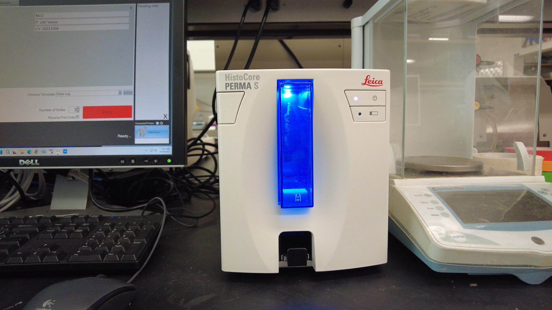
The sharp printing feature of the PERMA S significantly improves slide readability and helps reduce specimen misidentification. This slide printer enables individual slide printing at any microtome workstation. It also includes a 100-slide cartridge, facilitating swift and effortless replacement of slides.
News
Core Research Facilities proudly welcomed a remarkable addition: The Sigray QuantumLeap H2000 X-ray absorption spectroscopy equipment
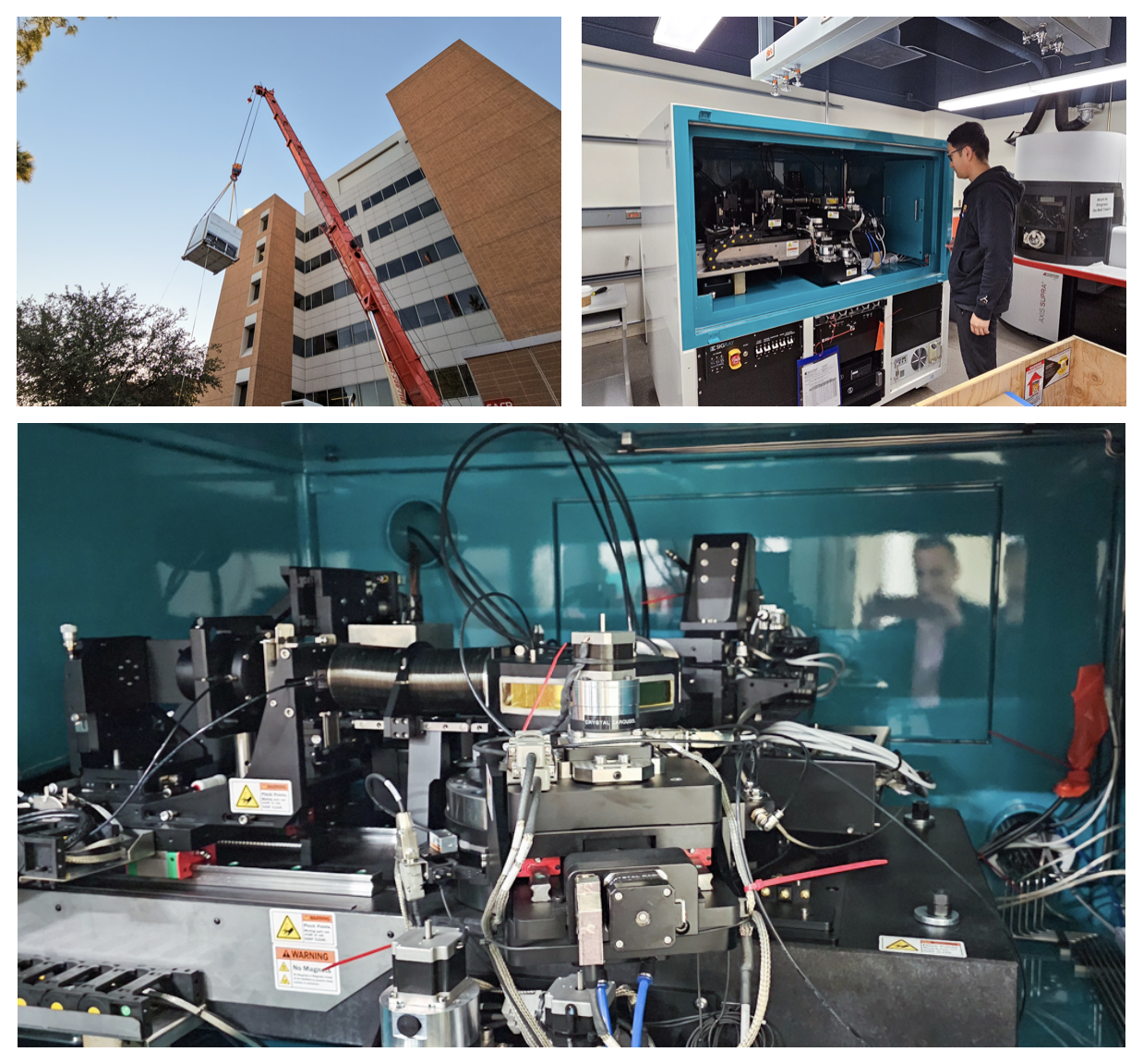
Funded by the New Economy Initiative: Advanced Materials, Processes and Energy Devices (AMPED) Science and Technology Center (STC) with Professor Mariana Bertoni as the lead researcher.
View the installation photos here!
Publications
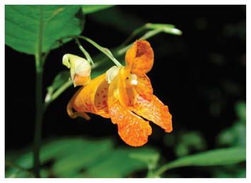
De-novno whole genome assembly of the orange jewelweed, Impatiens capensis Meerb. (Balsaminaceae) using nanopore long-read sequencing
Abstract
Researchers study the Balsaminaceae family, comprising two genera: Impatiens, with over 1,000 species, and Hydrocera, limited to the single species H. triflora. While Impatiens species are found across the Old World and North America, H. triflora is restricted to wetlands in South India, Sri Lanka and Southeast Asia.
Method
Plant tissue samples from Missouri were sent to Dovetail Genomics in California for DNA extraction and sequencing. The MinION device generated 3,311,362 sequencing reads, now accessible on GenBank in a public database.
Results
Researchers employed nanopore long-read sequencing for a high-quality Impatiens capensis genome. This supports a database for studying species diversity and evolution in Impatiens and Hydrocera, and aids in developing disease-resistant ornamental Impatiens.
Authors: Sudhindra R. Gadagkar, J. Antonio Baeza, Kristina Buss, Nate Johnson
Read the full research article.