Welcome to the ASU Core Facilities Newsletter. We are ready to support all your research goals. Please follow our LinkedIn page for additional resources and community information.
Regenerative Medicine Core Showcase
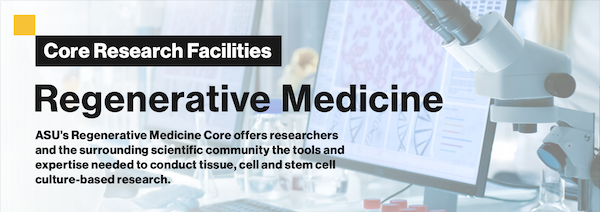
For this month's newsletter, we're going to focus on the people that make our Regenerative Medicine Core so great! Part of our larger Biosciences Core, the "Regen Med" facilities offer an extensive range of cell culture instruments, support and services.
The Regenerative Medicine Core houses the Histology, Flow Cytometry and Advanced Light Microscopy Cores, which makes it a comprehensive suite for medical research. The Core is equipped with state-of-the-art tools to support a wide array of research activities.
The Regenerative Medicine Core Facility at Arizona State University combines expertise on cellular and molecular biology, with the combination of microscopy, histology and flow cytometry.
The Regenerative Medicine Core facilitates research by offering training in cell and stem cell culture, nucleic acid analysis, biomolecule spectral analysis and techniques for preparing cells and tissues for analysis and sorting.
Regen Med provides advanced resources for cell and tissue imaging, including fully automated stations for tissue processing, sectioning, and staining, as well as sophisticated microscopes designed for both live and fixed sample imaging. This includes high-resolution microscopy, whole slide imaging capabilities, and comprehensive software packages for the analysis of complex images.
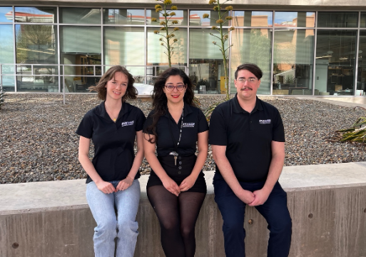
Meet the Regen Med team

Solange Steadman, the Facility Manager of our Histology Core, earned her biology degrees from ASU, including a recent master's. Her expertise in histology services for fixed tissues and user training has greatly enriched the capabilities of the new Histology Core within the Bioscience Core.
Adam Kindelin oversees the Flow Cytometry Core. He combines his biology degree from ASU with extensive neuroscience expertise gained at the Barrow Neurological Institute, specializing in fluorescently activated cell sorting (FACS), cell analysis, multi-color panel design, wet lab support and data analysis.
Our team provides advanced imaging instrumentation, trainings and services with the Advanced Light Microscopy Core.
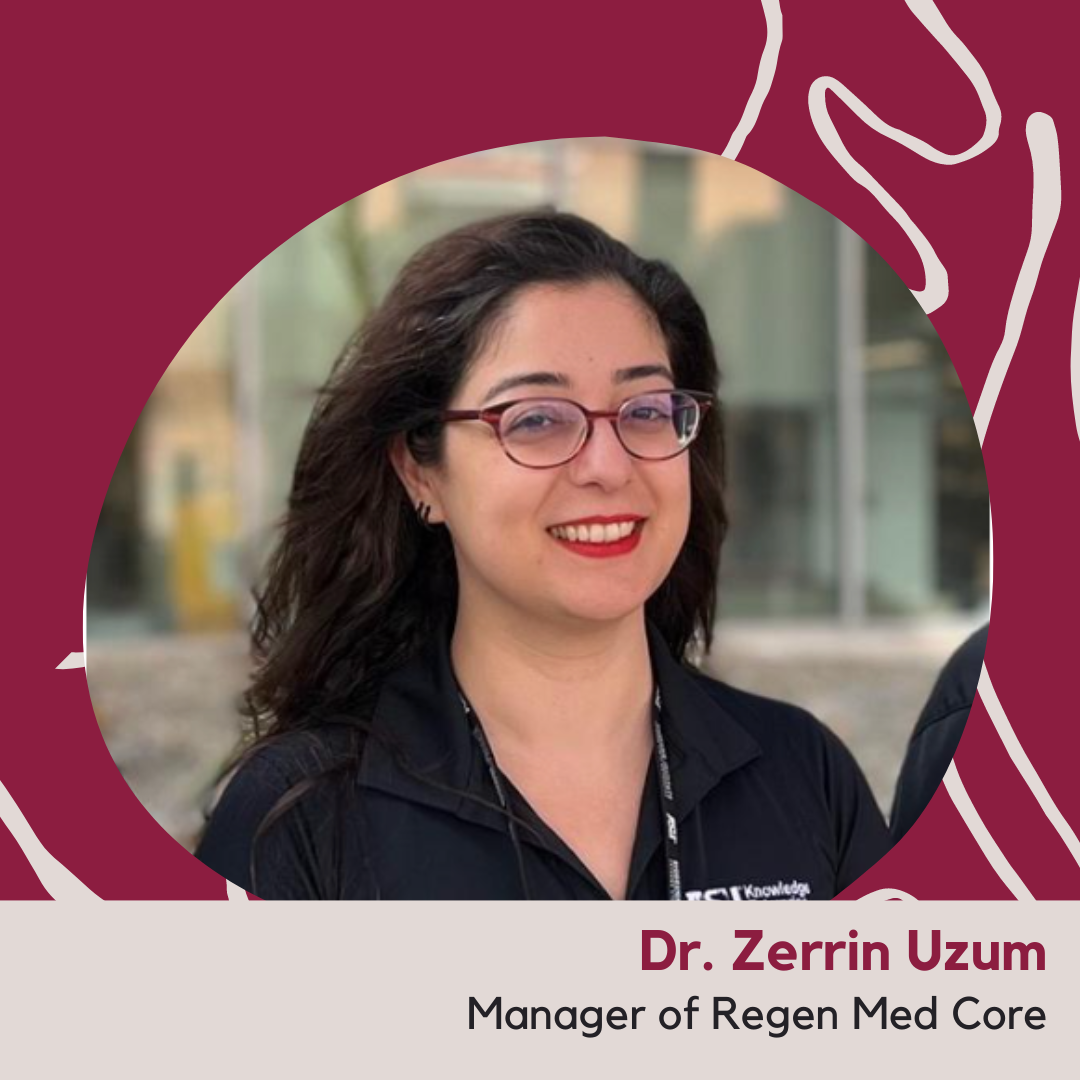
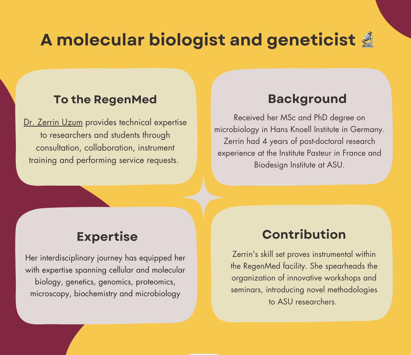
Our talented microscopists, Dr. Zerrin Uzum, Dr. Page Baluch and Dr. Honor Glenn , operate state-of-the-art microscopes, manage the cell culture facilities supporting the live imaging and train the student to acquire high quality images.
How Regen Med supports research.
News
Regenerative Medicine Educational Resources
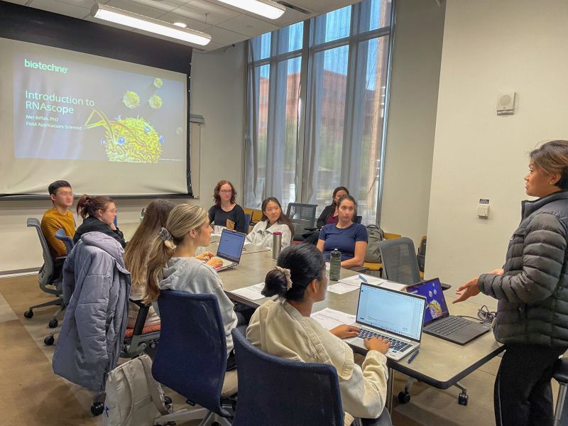
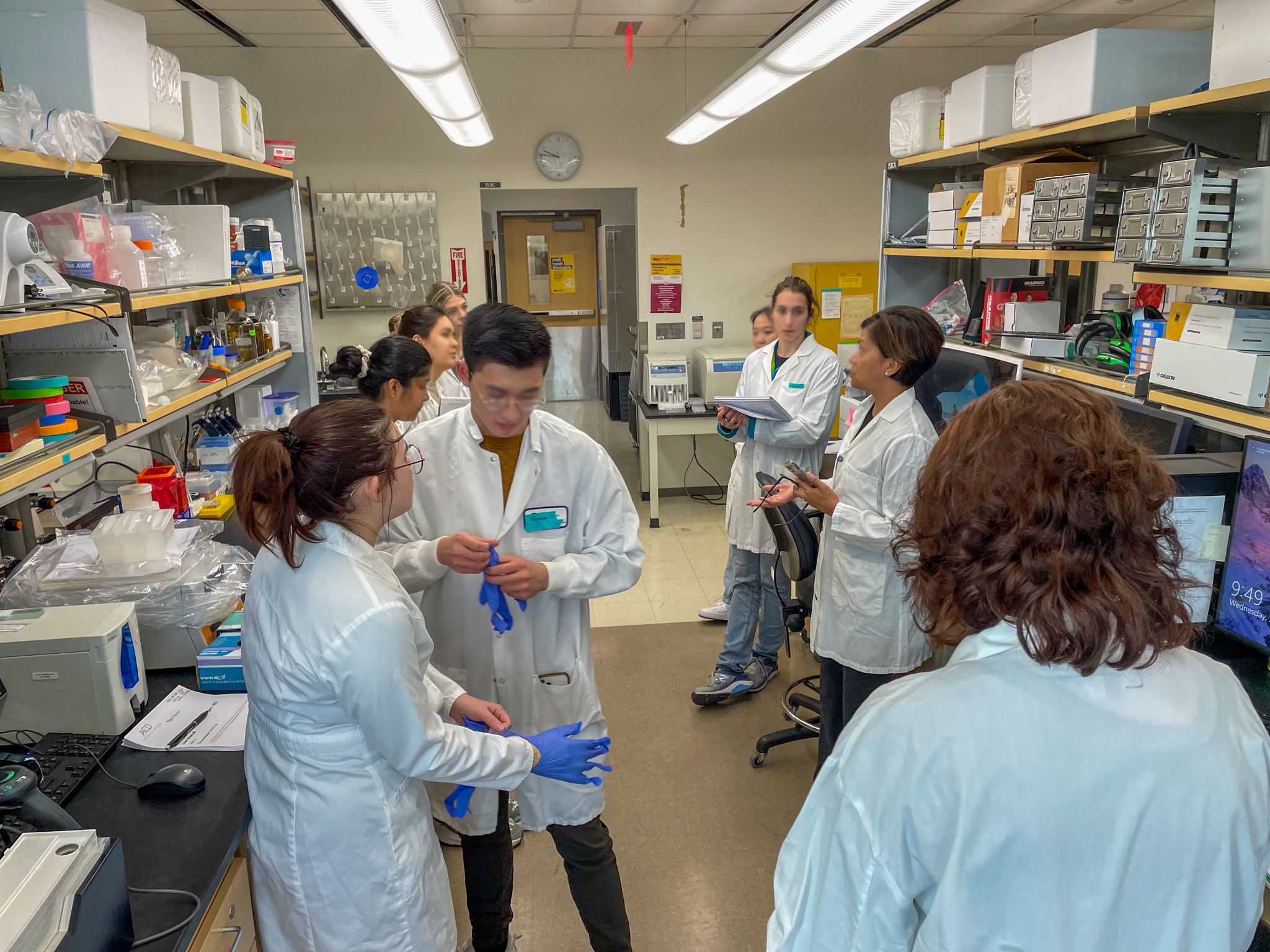
The Regen Med Core offers ASU scientists and students a variety of learning opportunities, including training on equipment and techniques from basic to advanced levels, technology demonstrations, hands-on workshops on innovative technologies, and seminars covering both fundamental concepts and the latest industry developments.
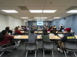
Regenerative Medicine Workshops and Seminars.
Regen Med Core Community Outreach
Dr. Page Baluch, Assistant Director of the Biosciences Core, manages multiple facilities, conducts research and champions science education for the wider community and youth.
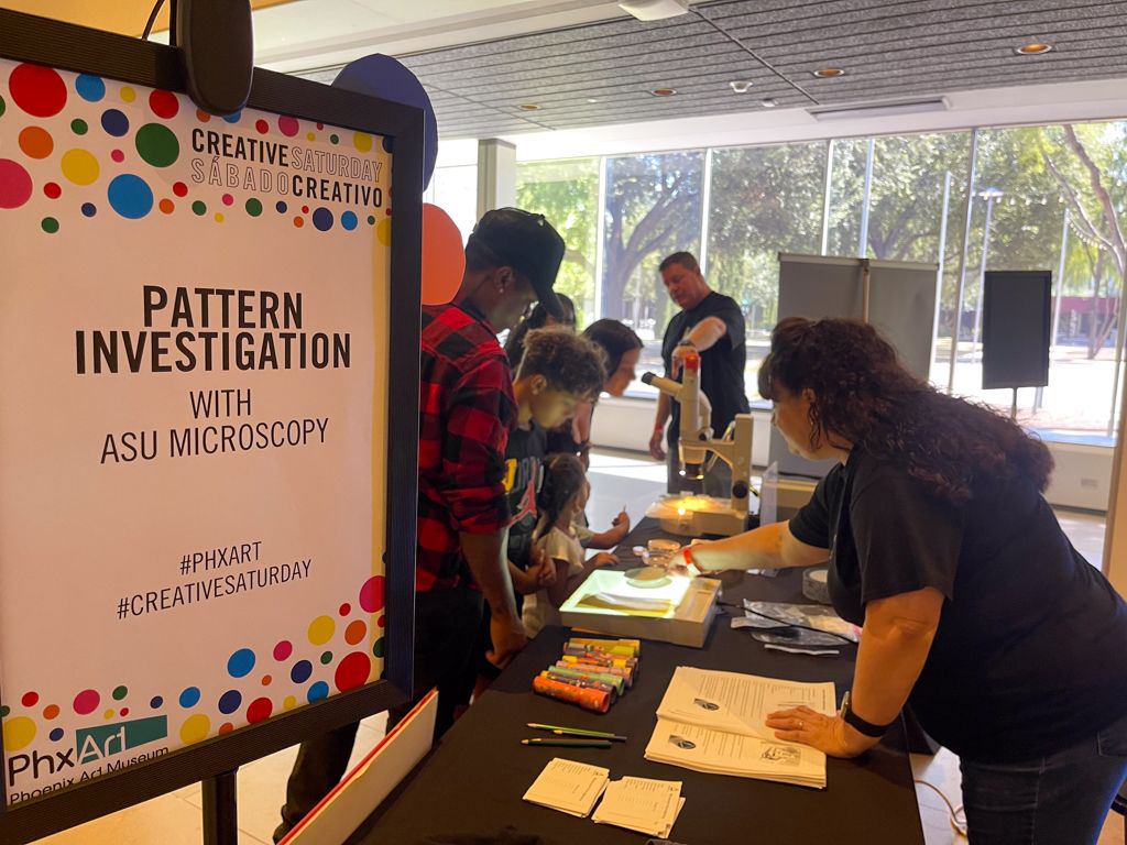
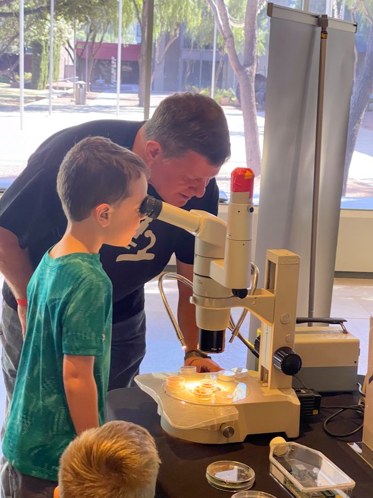
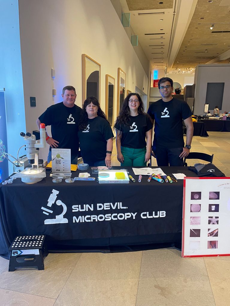
Dr. Baluch actively volunteers for editorial tasks for publications such as "Microscopy Today" and leads community outreach initiatives. She can often be found participating in Sun Devil Microscopy Club events. One example is the "Pattern Investigation with ASU Microscopy" booth she and Dr. Zerrin Uzum worked at a Phoenix Art Museum exhibition.
Publications
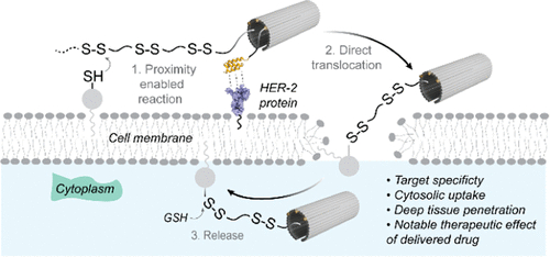
CytoDirect: A Nucleic Acid Nanodevice for Specific and Efficient Delivery of Functional Payloads to the Cytoplasm
This groundbreaking research utilized our Bioscience Core's Mass Spectrometry, Flow Cytometry and Advanced Light Microscopy facilities, showcasing their impact on global scientific advancements.
Abstract
CytoDirect is a DNA nanodevice that utilizes the programmability of DNA nanotechnology for efficient delivery directly into cancer cells, bypassing endo/lysosomal capture. This research underscores the effectiveness of affibody modifications, enhancing the capabilities of DNA nanostructures in targeted therapy and disease management.
Method
The study focuses on the design, construction, and analysis of CytoDirect, particularly its ability to target HER2-overexpressing SK-BR-3 breast cancer cells. It examines how CytoDirect is distributed within cells, investigates how it enters cells and explores its potential for delivering therapeutic oligonucleotides and small-molecule anticancer drugs.
Results
CytoDirect utilizes disulfide and HER2 affibody modifications on DNA origami for rapid cytosolic uptake and effective tumor penetration, significantly improving treatment in HER2-positive breast cancer. This method addresses the challenges of precise and effective drug delivery to deep tissues, offering a promising approach for enhanced therapy.
Delve into the researchers' findings.