Wide selection of equipment designed to prepare biological samples for electron microscopy imaging.
Balzers Critical Point Dryer
Critical-point drying is most commonly used with biological specimens destined for observations in the scanning electron microscope. The Balzers CPD020 utilizes liquid carbon dioxide, which has a critical-point at 31C and 73 bars pressure, in an enclosed compartment to circumvent ambient air-drying of the specimen. Critical-point drying minimizes structural changes and damage to specimen outer surfaces that occur during a normal liquid-to-gas phase change (i.e, air-drying) across the specimen surface
Leica Ice HPF
High-pressure freezing is used to preserve biological samples in their native state in millisecond time-frames and can provide superior results over traditional chemical-fixation techniques for EM analysis. By applying high-pressure (>30,000 PSI) in synchrony with a jet-spray of liquid nitrogen, water molecules within cells do not form large crystalline lattices, and samples up to approximately 1 mm diameter can be well-frozen without the damaging effects of ice crystal formation. After freezing, samples are subjected to further processing in preparation for EM. The Leica Ice can accommodate a variety of samples, including cell suspensions and adherent cells grown on sapphire disks.
Leica AFS
The Leica AFS performs freeze-substitution of high-pressure frozen samples and can gradually warm, cool or maintain a set temperature for samples according to a programmed schedule. The AFS can exchange solvents, perform additional fixation steps and embed samples before polymerizing resins under UV light at low temperatures for both high-pressure frozen and chemically fixed samples according to a user’s specific needs. Samples can be kept under gaseous LN2 to prevent contamination from moisture or oxygen.
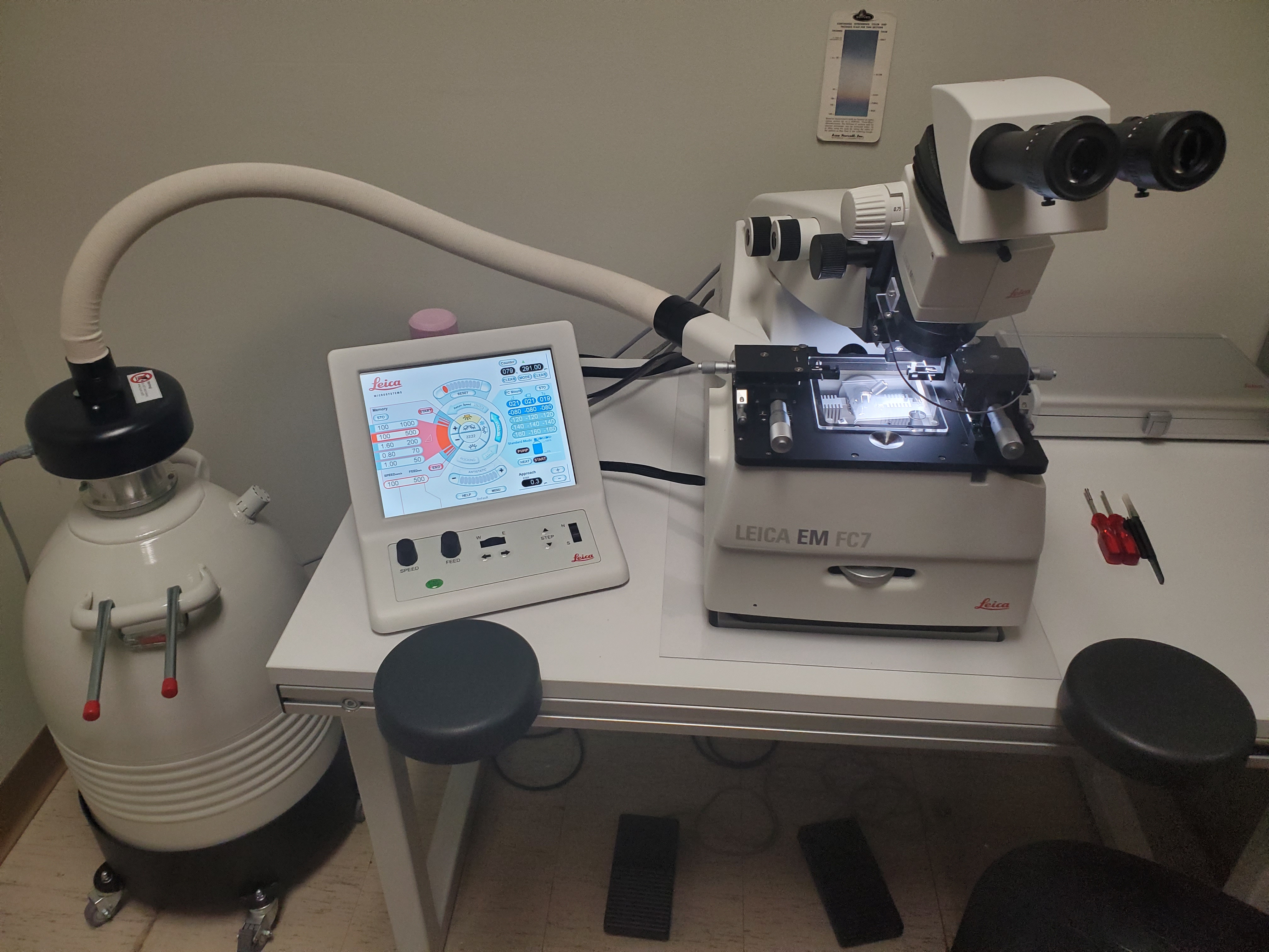
Microtomes
- Leica EM FC 7 Cryo Ultramicrotome
Designed for sectioning at low temperature for the preparation of TEM, SEM and AFM samples.
- Leica Ultracut R microtome
Designed for nanometer-scale sectioning of resin embedded biological samples destined for TEM analysis. The Ultracut-R has tri-directional illumination for viewing and ease of positioning sample blocks. It has a cutting range of 25-5000 nm.
- Reichert Ultracut E microtome
Designed for nanometer-scale sectioning of resin embedded biological samples destined for TEM analysis. The Ultracut-E has a cutting range of 25-990 nm.
Millrock Technology BT-48 Freeze Dryer
Freeze drying provides controlled desiccation of frozen samples at low pressure via sublimation of water content. The Millrock BT-48 unit has a 6-port manifold with various chamber volumes to accommodate a wide range of samples, and is capable of producing a condenser temperature of approximately -50C
Contact
- Sample preparation
| Cost for ASU Internal | Cost for ASU Internal with Staff Assistance | Cost for Other Academic/Non-Profit | Cost for Other Academic/Non-Profit with Staff Assistance |
|---|---|---|---|
|
Contact us for rates
|
Contact us for rates
|
Contact us for rates
|
Contact us for rates
|