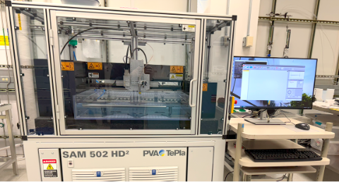
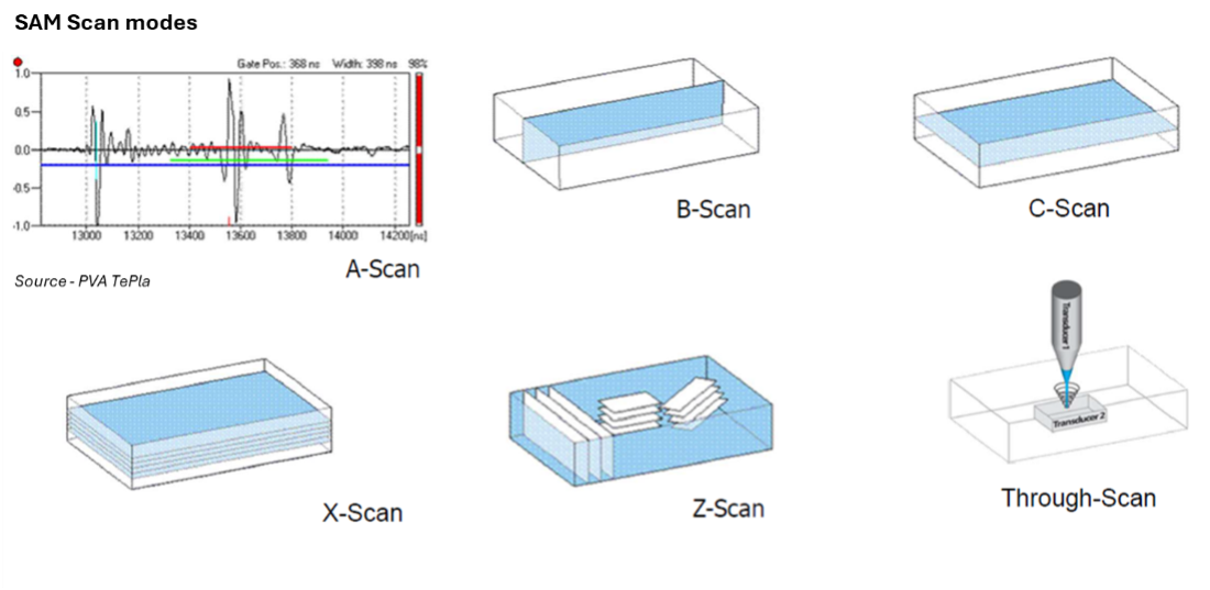
The scanning acoustic microscopy (SAM) uses high-frequency sound waves to image the underlying and internal structures of a sample. In typical operation, the sample is immersed in a coupling medium (usually water), where it is exposed to high-frequency acoustic waves produced by a piezoelectric transducer. The echoes generated by differences in acoustic impedance at material interfaces are then detected and analyzed to create images. Different imaging modes can be used to get detailed insights of layers and structures of components for internal voids, cracks and delamination at resolutions equivalent to that of a light microscope.

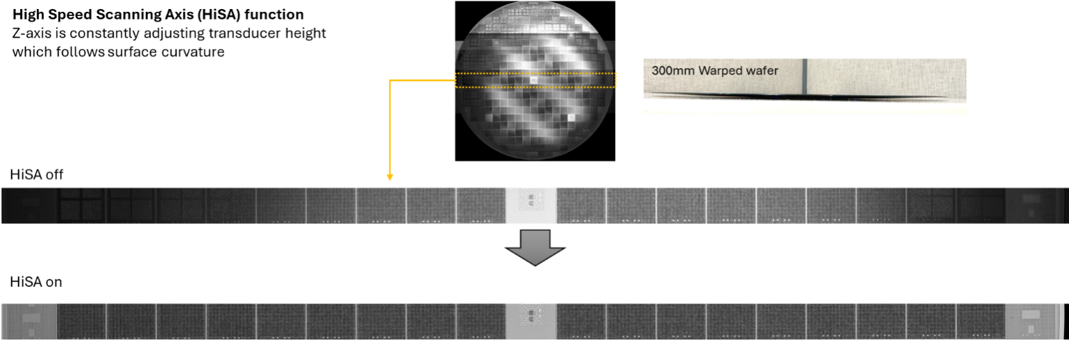
The SAM502 HD2 features HiSA (High Speed Scanning Axis) mode, which continuously tracks sample curvature during scanning to maintain optimal focus.
Sample requirements
- Solid phase
- Samples must be structurally and chemically stable in deionized water
Strengths
- It is a non-destructive and non-invasive imaging technique for internal defect detection
- Wide frequency range of transducers and focal lengths are available for a broad variety of samples
- Suitable for a wide range of sample sizes (Scanning range up to ~500 x 500mm)
Limitations
- Samples will be submerged in water during the imaging process
- Higher-frequency transducers provide better resolution but have limited penetration depth in thicker samples
- Non-planar or highly irregular sample shapes reduce imaging resolution, particularly in soft, porous, or rough materials (due to inconsistent scattering of sound waves)
- FileCSAM_112525.pptx10.42 MB
| Service | ASU rate | Nonprofit/other academic rate | Notes |
|---|---|---|---|
| Usage | $39/hr | $55/hr |